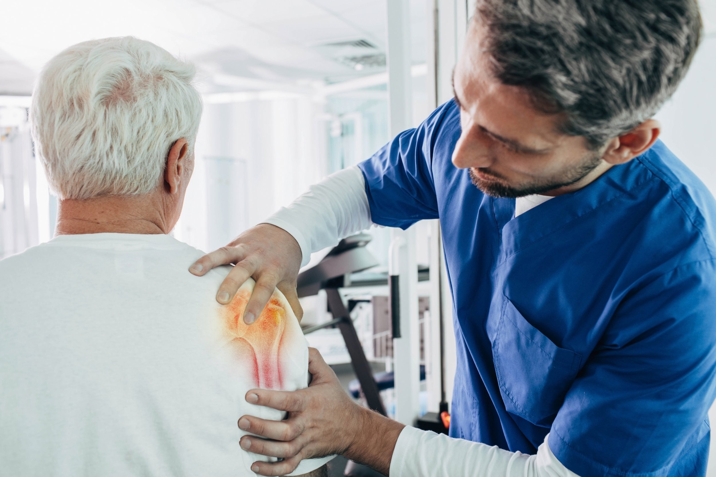
Normal Anatomy of the Shoulder Joint
The shoulder joint is a complex and highly mobile joint that enables a wide range of movements, making it one of the most flexible joints in the human body. It is formed by the articulation of three bones: the humerus (upper arm bone), the scapula (shoulder blade), and the clavicle (collarbone). The shoulder joint consists of several structures that work in harmony to facilitate movement and provide stability. Here’s a detailed overview of the normal anatomy of the shoulder joint:

Bones:
- Humerus: The upper arm bone with a rounded head that fits into the shallow socket of the scapula, forming the glenohumeral joint, which is the primary joint of the shoulder.
- Scapula (Shoulder Blade): A flat, triangular bone that articulates with the humerus and the clavicle. The scapula provides attachment sites for various muscles that contribute to shoulder movement.
- Clavicle (Collarbone): A slender bone that connects the sternum (breastbone) to the scapula, providing stability to the shoulder joint and allowing it to move away from the midline of the body.
Joints and Articulations:
- Glenohumeral Joint: Also known as the shoulder joint, it’s a ball-and-socket joint formed by the head of the humerus and the shallow glenoid cavity of the scapula. This joint allows a wide range of motion, including flexion, extension, abduction, adduction, internal rotation, and external rotation.
- Acromioclavicular (AC) Joint: Located where the clavicle meets the acromion (a bony projection of the scapula), this joint allows limited movement and provides stability to the shoulder complex.
- Sternoclavicular (SC) Joint: The joint where the clavicle connects to the sternum. It allows limited movement during shoulder elevation and protraction/retraction.
Muscles and Tendons:
- Rotator Cuff Muscles: A group of four muscles (supraspinatus, infraspinatus, teres minor, and subscapularis) that surround the shoulder joint, stabilizing it and facilitating rotation and abduction of the arm.
- Deltoid Muscle: A large muscle covering the shoulder, responsible for lifting the arm and providing its shape.
- Biceps and Triceps Muscles: These muscles attach to the humerus and contribute to various shoulder movements.
Ligaments:
- Glenohumeral Ligaments: Ligaments that help stabilize the glenohumeral joint by connecting the humerus to the glenoid cavity of the scapula.
- Coracoclavicular Ligaments: These ligaments help stabilize the AC joint.
Labrum:
The labrum is a ring of fibrous cartilage that surrounds the glenoid cavity, deepening the socket and providing stability to the glenohumeral joint.
Bursae:
Bursae are fluid-filled sacs that reduce friction between tendons, ligaments, and bones. In the shoulder joint, bursae help protect and cushion structures during movement.
Nerves and Blood Supply:
Various nerves (e.g., brachial plexus) and blood vessels supply the shoulder joint and its surrounding structures, ensuring proper innervation and circulation.
Conclusion:
The shoulder joint’s intricate anatomy enables a remarkable range of motion and functionality. Understanding the normal structures and their interactions within the shoulder joint is essential for diagnosing and treating shoulder injuries or conditions and for maintaining optimal shoulder health. If you experience any shoulder pain, discomfort, or limitations in movement, consulting a healthcare professional can provide insights into your shoulder’s condition and guide appropriate treatment.


